Figure 2.

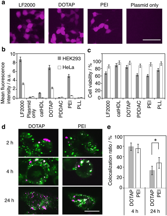
Intracellular gene delivery by cationic AuNRs. (a) Fluorescence images of HEK293T cells treated with cationic AuNR/pCMV-DsRed complexes. LF2000 was used as a positive control. Scale bar = 100 µm. (b) Transfection efficiency determined by flow cytometry analysis. Data indicate the mean fluorescence intensity of DsRed (n = 3, average ± SD). HEK293T cells and HeLa cells were treated with AuNR/pCMV-DsRed complexes ([Au] = 20 µg/mL, pCMV-DsRed = 2 µg/mL) for 24 h. DOTAP-AuNRs show a high transfection efficiency, comparable to that of LF2000. (c) Cell Count Kit-8 (CCK-8) assay data for HEK293T cells and HeLa cells treated with AuNR/pCMV-DsRed complexes for 24 h. Data indicate the mean cell viability (n = 3, average ± SD). The cytotoxicity of cells treated with DOTAP-AuNRs is lower than that of those treated with LF2000. (d) Time-dependent change of the localization of AuNRs in HEK293T cells. DOTAP- and PEI-AuNRs were labeled with Rho-PE and Alexa Fluor 546, respectively. AuNR localization (red signal) was analyzed after 2, 4 and 24 h treatment. Late endosomes/lysosomes were stained with LysoTracker Green DND-26 (LysoTracker, green). (e) The colocalization ratios of the red pixels to green pixels after 4 and 24 h were calculated, respectively (n = 10, average ± SD). Asterisks indicate P values < 0.05 determined using Student’s t-test. Scale bar = 10 µm.
