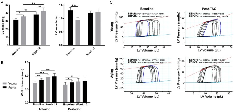
Figure 3.
Myocardial hypertrophy post-TAC. At baseline, aging mice exhibit greater heart weight, LV mass, anterior wall thickness and posterior wall thickness. Young and aging mice undergo significant hypertrophy 12 weeks post-TAC (A). Heart weight, anterior and posterior wall thickness of young and aging mice are no longer discernibly different between young and aging mice 12 weeks post-TAC (B). Baseline and 12-week post TAC values were compared using paired t-tests, and values were compared across groups at each time-point using non-paired t-tests. (C) Representative pressure volume loops from young and aging mice. *P<0.05, **P<0.01, ***P<0.001 (n=6 young mice, n=6 aging mice).

