
Fig. 8.
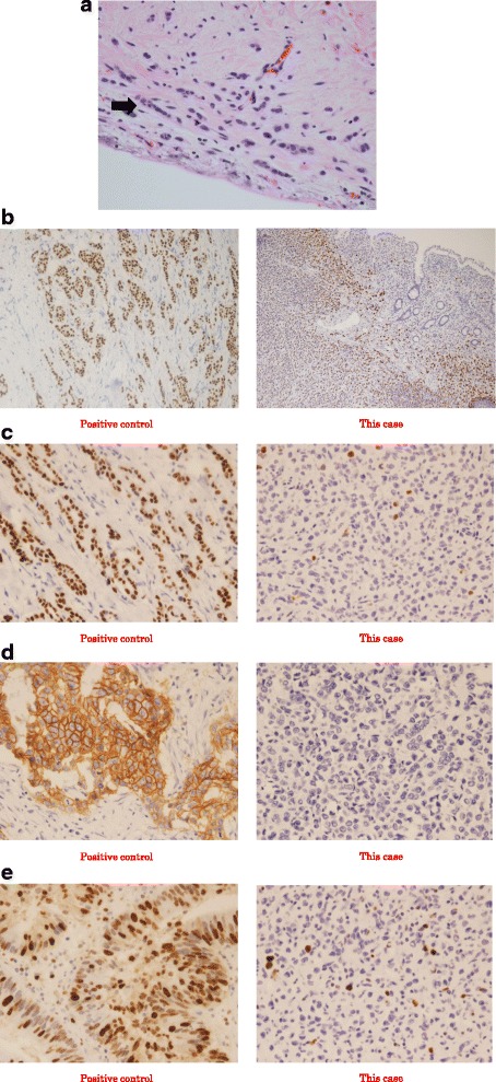
Histological examination. The findings of histological examination indicate invasive lobular carcinoma. a Hematoxylin and eosin staining of the tumor (×400). b Estrogen receptor (ER) staining of the tumor (×200). The positive rate of ER was 100%. c Progesterone receptor (PgR) staining of the tumor (×400). The positive rate of PgR was 2–3%. d Human epidermal growth factor receptor 2 (Her-2) staining of the tumor (×400). The positive rate of Her-2 was 0%. E Ki-67 staining of the tumor (×400). The positive rate of Ki-67 was 5–10%