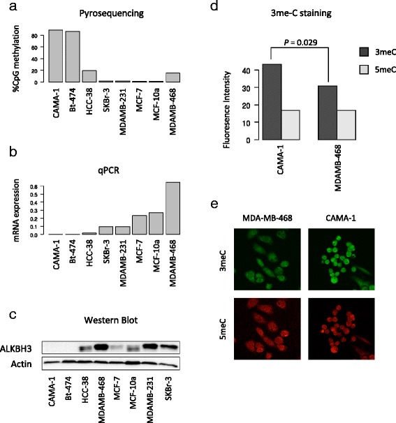
Fig. 3.

Epigenetic silencing of ALKBH3 and accumulation of 3-methylcytosine damage. a CpG methylation analysis for the ALKBH3 gene promoter by pyrosequencing and (b) ALKBH3 mRNA expression analysis by qPCR in breast cancer cell lines (CAMA1, Bt-474, HCC-38, SKBr-3, MDA-MB-231, MCF-7 and MDA-MB-468) and a mammary epithelial cell line derived from a fibrocystic lesion of the breast (MCF10A). The association between ALKBH3 promoter methylation and mRNA expression was found to be statistically significant (Spearman’s rho = −0.73; P = 0.039). c ALKBH3 protein expression analysed by western blotting using the same panel of breast-derived cell lines revealing lack of expression in two cell lines (CAMA-1 and Bt-474). Actin protein expression is shown for comparison. d Immunofluorescent staining for 3-methylcytosine and 5-methylcytosine in CAMA-1 (ALKBH3 methylated) and MDA-MB-468 (ALKBH3 unmethylated). The P-value shown was derived from Wilcoxon’s rank sum testing for differences between CAMA-1 and MDA-MB-468 with respect to intensity for 3-methylcytosine. As expected, no statistically significant differences were found with respect to overall 5-methylcytosine intensity levels between CAMA-1 and MDA-MB-468. e Representative images showing 3-methylcytosine (green) and 5-methylcytosine (red) fluorescence staining in CAMA-1 and MDA-MB-468
