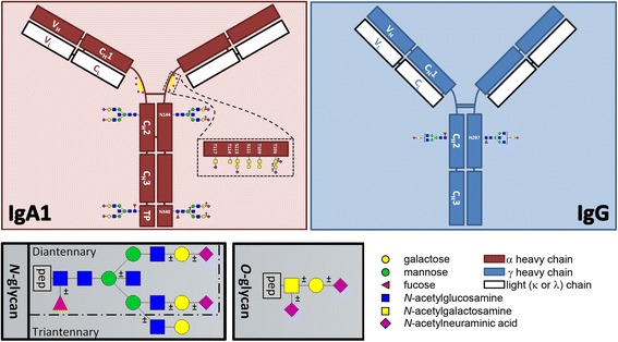
Fig. 1.

Schematic representation of immunoglobulin A (IgA) and IgG and the sites of their respective glycosylation sites as can be detected by glycopeptide analysis of serum-derived samples. Insets show schematic representations of an O-glycan, an N-glycan and a potential configuration of an O-glycopeptide. No linkage information is intended by any position of a monosaccharide
