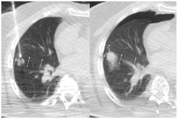
Figure 3. Example CPLB case with transfissural needle path.

Patient with history of colon cancer underwent CPLB of a new right lower lobe lesion with an anterior approach through the right middle lobe. Intraprocedural CT fluoroscopy image (left) with arrows indicating a non-vascular line hyperdense to the lung parenchyma corresponding to the right major fissure. Immediate post biopsy CT fluoroscopic image demonstrates a small anterior pneumothorax. The needle tract is indicated with a dotted arrow. This patient ultimately required a chest tube for just over 3 days.
