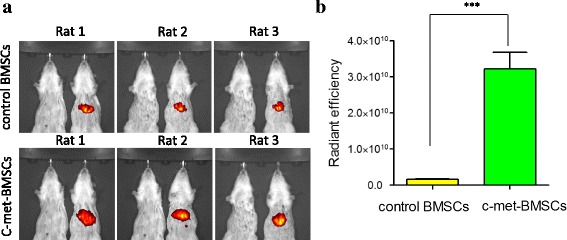
Fig. 4.

Analysis of cell migration in ALF rats transplanted with c-Met-BMSCs using an in vivo imaging system. DiR dye was used to label the c-Met-BMSCs and control BMSCs. The same amount of c-Met-BMSCs and control BMSCs were transplanted into ALF rats through the vena caudalis. After 24 h cells that had migrated to the injured liver were detected by an imaging system, and the fluorescent intensity was measured. a The fluorescent intensity of the liver tissues in rats transplanted with control bone marrow-derived mesenchymal stem cells (BMSCs) or c-Met-BMSCs. b The difference between the two groups was statistically significant. Data are presented as mean ± SD. ***P < 0.001
