Figure 3.
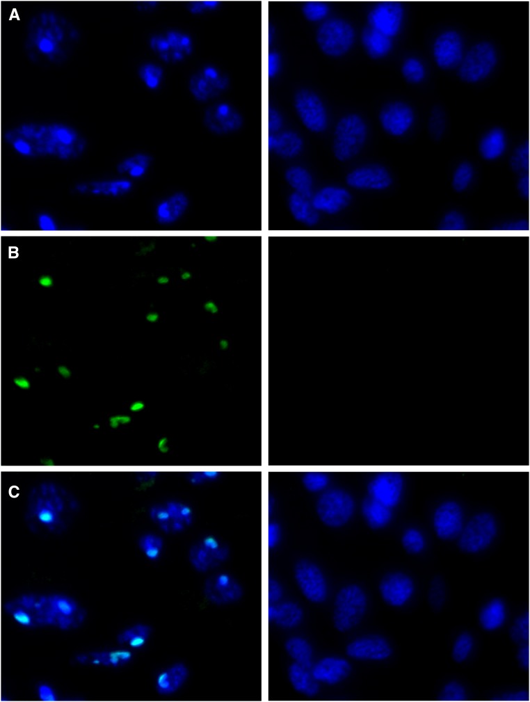
DAPI (A), H3K9me3 (B), and merged (C) images of male (left panels) and female (right panels) Liposcelis sp. abdominal tissue. Condensed regions of DAPI staining that colocalize with H3K9me3 staining are present in male tissue but absent from female tissue, indicating chromocenters are present in male cells. Bar, 5 μm.

