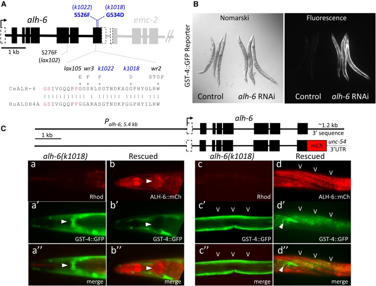
Figure 1.
Identification of xrep-2 as an allele of alh-6. (A) The alh-6 gene structure and mutations. The gene structure of alh-6 is diagramed (black) along with part of its neighboring gene in the operon, emc-2 (gray). The positions of alleles identified in this study (k1018 and k1022) are shown in blue relative to several previously identified mutations [Schlipalius et al. (2012) and Pang and Curran (2014)]. Many cluster in the last exon, which encodes an evolutionarily conserved interface between the inferred substrate and NAD+-binding pockets of ALH-6 based on sequence homology to mammalian ALDH4A1 (Srivastava et al. 2012). A segment of the protein sequence from this region is shown, with active site residues in red and mutant substitutions (black and blue) as indicated. (B) Phenocopy of the xrep-2 mutation by alh-6 RNAi (RNA interference). Control wild-type adult animals harboring the gst-4::gfp translational fusion reporter gene are shown next to the same strain after alh-6 RNAi. Knockdown of alh-6 activity results in strong upregulation of gst-4::gfp expression in bodywall muscles (BWMs). (C) alh-6 mutant rescue. Genomic wild-type and mCherry (mCh)-tagged alh-6-rescuing constructs are diagramed at the top; each was introduced separately into alh-6(k1018) mutants harboring the gst-4::gfp reporter gene and stable extrachromosomal strains were established. The left panels (a and b series) illustrate the head expression, emphasizing pharyngeal patterns for both reporters; the arrowhead indicates expression in the posterior pharyngeal bulb. Note that expression of the mCh-tagged wild-type alh-6 transgene is strong in the posterior bulb of the pharynx (b and b”; arrowhead) and completely suppresses the mutant pattern of gst-4::gfp expression in this tissue (a’ and a”; arrowhead). A similar comparison of mutant and rescue transgene expression is shown for midbody BWMs and hypodermal cells (HYPs) (c and d series), with BWM expression of alh-6::mCh (carets) suppressing gst-4::gfp expression. The arrowhead (d’ and d”) points to a single, nonrescued BWM cell still expressing gst-4::gfp; background gut auto-fluorescence captured in the GFP channel is also visible in these images.

