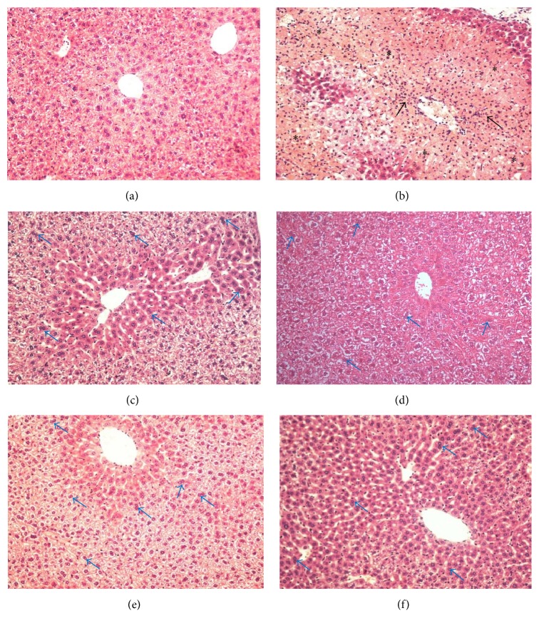
Figure 5.
Effect of pretreatment with citral on the liver tissue morphology. (a) Control mice liver showed normal morphology and absent lesion area; (b) APAP group (mice liver that received orally APAP on last day of treatment, 250 mg/kg): presence of severe necrosis (∗) and inflammatory infiltrate (arrows); (c) group pretreated with SLM (200 mg/kg) + APAP; (d) 125 mg/kg citral + APAP; (e) 250 mg/kg citral + APAP; (f) 500 mg/kg citral + APAP. ((c)–(f)) Presence of binucleate hepatocytes (arrows in blue) and mild lesion area. Original magnification 20x. The sections stained with hematoxylin and eosin.

