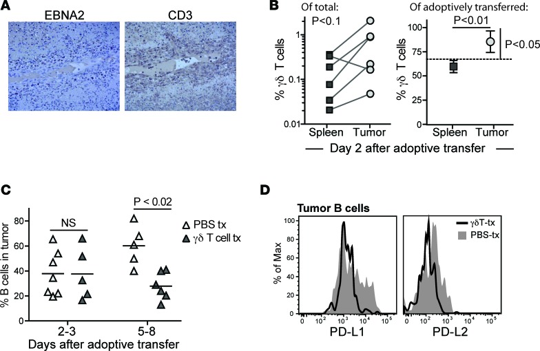
Figure 8. Recruitment of γδ T cells and analysis of tumor B cells after late time point γδ T cell administration.
(A) Light microscopic images (×10 magnification) of IHC of serial sections of tumor tissue from a γδ T cell–treated mouse showing cells expressing the viral protein EBNA2 (left image) or CD3 (right image). T cells are observed within the blood vessel in the middle of the field, as well as infiltrating the surrounding tissue in areas where EBNA2+ cells are localized. (B) Adoptively transferred γδ T cells appear slightly enriched in tumor tissue compared with spleen. Left plot: percentage of human T cells expressing Vδ2 in paired spleen versus tumor tissue samples from independent mice. Tissues were harvested 2 days after administration of γδ T cell immunotherapy. The P value was calculated using a paired, nonparametric t test (Wilcoxon signed-rank test). Right plot: percentage of the adoptively transferred cells in spleen versus tumor expressing a Vγ2Vδ2+ TCR. The dashed line shows the frequency of γδ T cells in the injected population; symbols show mean and standard deviations of 4 replicate analyses each of spleen and tumor tissue. The P value comparing spleen versus tumor frequencies (horizontal line) was calculated using an unpaired parametric t test; the P value comparing tumor mean to the γδ T cell frequency of the injected cells (vertical bar) was calculated using a 1-sample, 2-tailed t test. (C) B cells as a percentage of the total human cells in tumor tissue from a series of mice that were administered γδ T cells (dark triangles) or vehicle (light triangles). Tumors were harvested at the indicated times after adoptive transfer or mock treatment. The P value was calculated using a 2-tailed nonparametric t test (Mann-Whitney analysis). (D) Flow cytometric analysis of PD-L1 and PD-L2 staining on tumor B cells. Filled gray histograms show results from a mouse given vehicle (PBS-tx); black line shows results from a mouse given γδ T cells. tx, transfer.

