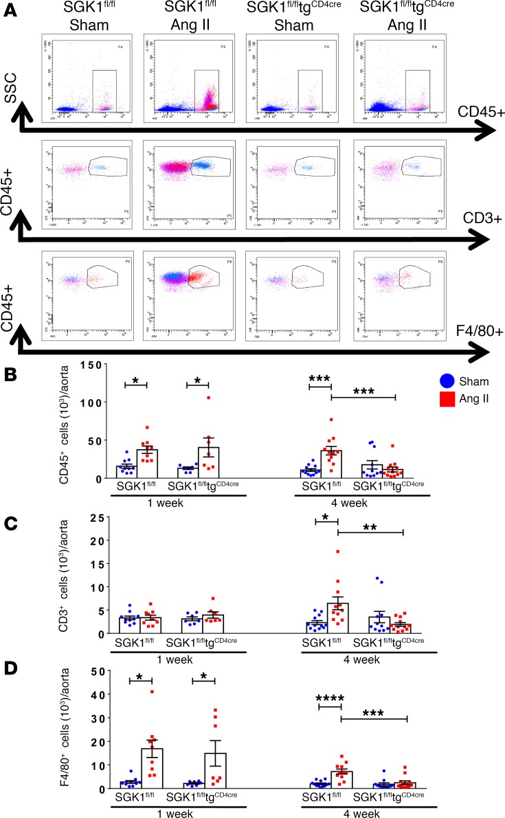
Figure 2. T cell SGK1 deficiency prevents the chronic phase of angiotensin II–induced vascular inflammation.
(A) Representative flow cytometry dot plots showing gating strategy for total leukocytes (CD45+ cells), total T lymphocytes (CD45+CD3+ cells), and monocytes/macrophages (CD45+F4/80+ cells) in single-cell suspensions from the thoracic aorta of SGK1fl/fl and SGK1fl/fltgCD4cre mice infused with angiotensin II (Ang II) or vehicle (sham) for 7 or 28 days. (B–D) Summary data of absolute numbers of indicated cell types per thoracic aorta. *P < 0.05, **P < 0.01, ***P < 0.001, ****P < 0.0001; 2-way ANOVA/Holm-Sidak’s post-hoc test; n = 7–12 per group. All data are expressed as mean ± SEM.

