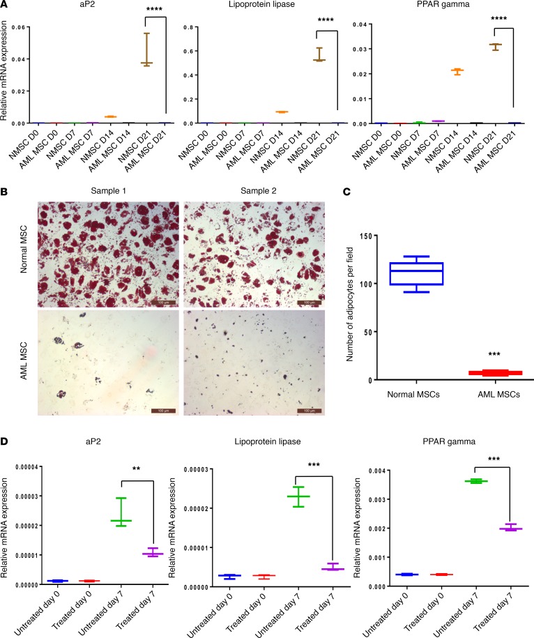
Figure 3. AML cells inhibit adipogenic differentiation in MSCs.
(A) N- and AML-MSCs were cultured in adipogenic differentiation medium for 3 weeks. mRNA expression of adipocyte-associated genes, aP2, lipoprotein lipase, and PPARγ, in N- and AML-MSCs before and after induction of differentiation was analyzed weekly by qRT-PCR. (B) N- or AML-MSCs (n = 5 each group) were cultured in adipogenic differentiation medium for 28 days. On day 28, the adipocytes were stained with Oil Red O. Scale bar: 100 μm. (C) Oil Red O–positive cells (i.e., adipocytes) were counted in 10 microscopic fields per sample. (D) N-MSCs were pretreated with OCI-AML3–conditioned medium for 5 days before they were cultured in adipogenic differentiation medium for 7 days. Expression of indicated adipogenic lineage–associated genes before induction of differentiation and on day 7 was analyzed by qRT-PCR. GAPDH served as an equal loading control. Two-way ANOVA was used for comparisons of 3 or more groups and unpaired Student’s t test was used for comparisons of 2 groups (**P < 0.01, ***P < 0.001. ****P < 0.0001 versus control). In addition, Tukey’s multiple comparison test was also performed for multiple data sets.

