Figure 1.

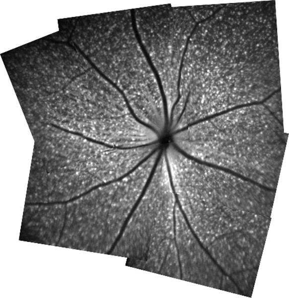
Retinal montage constructed from multiple in vivo blue-light confocal scanning laser ophthalmoscopy images. Each fluorescent spot represents Thy-1 expressing retinal ganglion cell.

Retinal montage constructed from multiple in vivo blue-light confocal scanning laser ophthalmoscopy images. Each fluorescent spot represents Thy-1 expressing retinal ganglion cell.