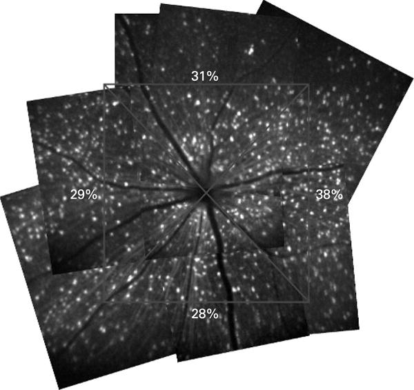
Figure 6.

Retinal montages showing diffuse loss of retinal ganglion cells (RGCs) after ischaemic reperfusion injury. The spatial distribution of RGC loss was examined with retinal montages constructed from multiple blue-light confocal scanning laser ophthalmoscope images covering a retinal area of approximately 2×2 mm2 with centre at the optic disc. The remaining proportions of RGCs in each quadrant are shown.
