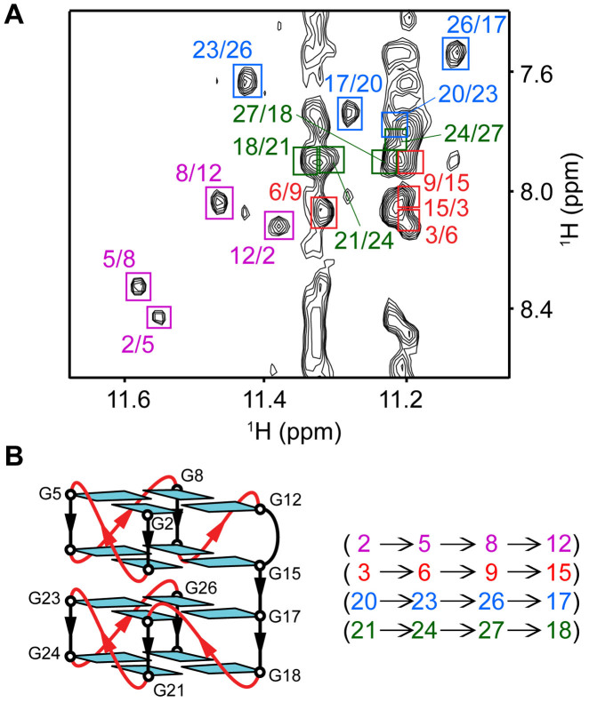
Figure 4.
Determination of G-quadruplex folding topology of AT11 in K+ solution. (A) NOESY spectrum (mixing time, 200 ms) showing the guanine H1-H8 NOE connectivity. Cross-peaks between the imino (H1) protons and aromatic (H8) protons are framed and labeled with the H1 and H8 proton assignment in the first and second position respectively. The first, second, third and fourth G-tetrad layers are colored in magenta, red, blue and forest green respectively. (B) Schematic structure showing the G-tetrad alignments and folding topology of AT11.

