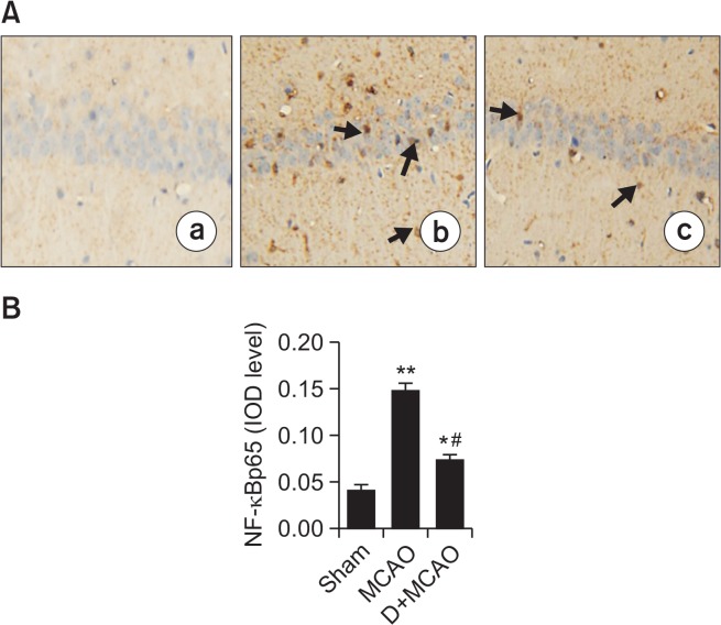
Fig. 4.
Effect of dexmedetomidine on hippocampal NF-κBp65 expression in MCAO rats. (A) Photomicrographs of NF-κBp65 immunoreactivity in hippocampal CA1 (200 amplification). There were slightly NF-κBp65-positive expression in Sham group (a), while in MCAO rats (b), abundant NF-κBp65-positive expression in cytoplasm and nucleus of hippocampal neurons with brown staining (arrow), whereas in D+MCAO group, positive expression of NF-κBp65 was decreased (c). (B) Quantification of NF-κBp65 IOD level. *p<0.05, **p<0.01 vs. Sham; #p<0.01 vs. MCAO.

