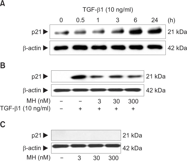
Fig. 2.
The effect of 4-O-Methylhonokiol (MH) on TGF-β1-induced p21 expression in HaCaT cell. (A) HaCaT cells were treated with TGF-β1 (10 ng/mL) for the indicated times. Whole lysates (30 μg proteins) were examined by 10% SDS-PAGE and analyzed with Western blotting using for p21. (B) After pretreatment with MH (3, 30 and 300 nM) for 1 h, HaCaT cells were treated with TGF-β1 (10 ng/mL) for another 24 h. The expression level of p21 was analyzed by Western blotting. (C) HaCaT cells were incubated with MH (3, 30 and 300 nM) for 24 h, and the expression level of p21 was analyzed. Blots were then probed with the corresponding antibodies after which they were stripped and reprobed with an antibody to β-actin to ensure equivalent loading and transfer.

