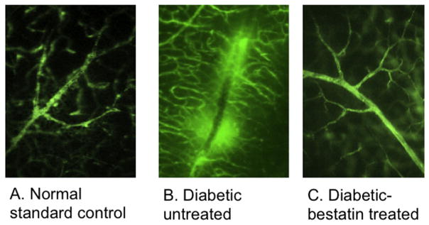
Fig. 1.
Following intravenous injection of BSA-FITC, flat mount retinas were visualized using fluorescence microscopy to determine the extent of FITC diffusion around retinal blood vessels. (A) No spot of hyperfluorescence was seen in normal non-diabetic mice retinas. (B) Diabetic mice retina had multiple patchy areas of hyperfluorescence (white arrows) (C) Intravitreal bestatin treated mice eyes did not reveal any spot of hyperfluorescence.

