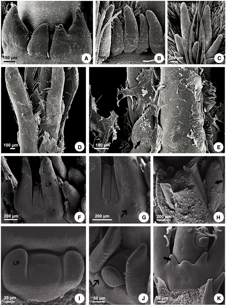
Figure 1.
Scanning electron microscopy of colleters in Apocynaceae. (A, D, E) Peplonia axillaris. (B) Asclepias curassavica. (C) Fischeria stellata. (F–H) Allamanda schottii. (I–K) Blepharadon bicuspidatum. (A) Stipular colleters. (B) Petiolar colleters. (C) Laminar colleters. (D) Colleters in the shoot apex. (E) Colleters secretion in the shoot apex. (F–K) Initiation of stipules from leaf primordia. (F, G, J) Projection of the stipules from leaf primordia base. (H) Colleters originated from stipules laterally to the petiole. (I) Leaf primordia without stipules. (K) Colleters origin from sheathing stipules. LP = leaf primordium. P = petiole. Narrow arrow = stipule. Large arrow = stipular colleter.

