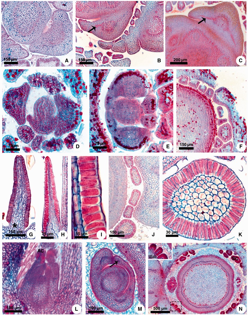
Figure 4.
Stipule development and colleter structure in Apocynaceae and Rubiaceae. (A) Secontatia densiflora. (B, C) Thevetia peruviana. (D) Mandevilla tenuifolia. (E, F) Secondatia densiflora. (G) Fisheria stellata. (H, I) Peplonia axillaris. (J, K) Asclepias curassavica. (L–N) Mussaenda erythrophylla. (A–F, J, K, M, N) Transversal sections. (G–I, L) Longitudinal sections. (A–C) Projection of the vascularized stipules as a pair laterally to the petiole. (D) Projection of the stipule from leaf primordium base. (E) Fusion of the sheathing stipules of a node. (F, N) Sheathing stipules giving rise to colleters. (G, H) General view of colleters. (I) Detail of the secretory epidermis. (J) Stipular and petiolar colleters. (K) Detail of the stipular colleter. (L) General view of the shoot apex. (M) Sheathing stipules with vascular bundles. (N) Detail of the stipular colleters (inset). * = colleter peduncle. Arrow = vascular bundle of the stipule. AC = apical colleter of the stipule. SC = stipular colleter.

