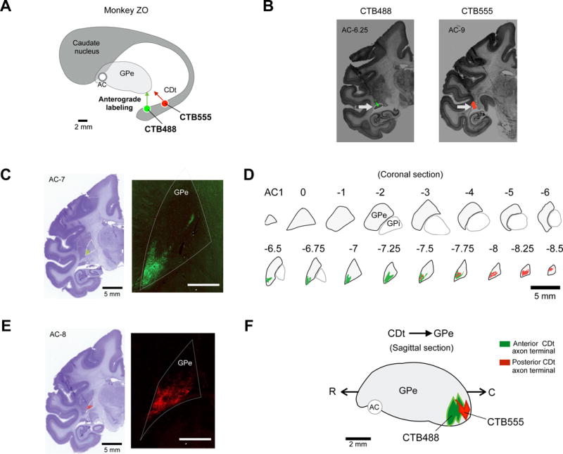
Figure 1. CDt projection to caudal-ventral region of GPe.

(A) Scheme of injection sites in CDt in monkey ZO. CTB488 and CTB555 were injected in anterior and posterior regions of CDt respectively. (B) Injection sites in the anterior and posterior CDt. Fluorescent signals were detected in each region of CDt (White arrows). (C and E) Examples of axon terminal signals in GPe. Axonal plexus (green and red signals) were detected in coronal slices of GPe. White bar: 5 mm (D) CDt projection sites in coronal slices of GPe. Axon terminals of CDt neurons were mainly found in caudal-ventral regions of GPe (cvGPe). (F) Sagittal view of CDt-projection site in GPe. CTB555 and CTB488 signals were topographically organized in rostral-caudal axis of GPe.
