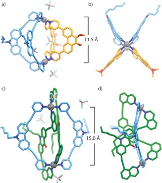
Figure 3.

X‐ray crystal structures of cis‐[Pd2 LC 2 LP 2]4+ (2) and trans‐[Pd2(anti‐LA)2 LC 2]4+ (3): a) the structure of 2 showing the occupation of the cavity by two BF4 − counterions; b) top view of 2; c) perspective view of 3 showing the ligand‐occupied cavity; d) side view of 3 showing the trans/anti arrangement of LA. Pd⋅⋅⋅Pd distances are shown and hydrogen atoms are removed for clarity.25
