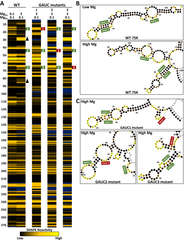
Figure 2.
SHAPE analysis of GAUC mutants. (A) SHAPE results for wild type (WT, from Figure 1) and the three GAUC to CUAG mutations (green numbered boxes) folded under the indicated magnesium folding conditions. (B) Secondary structure predictions of low and high magnesium folded 7SK using RNAstructure and corresponding SHAPE constraints and modeled with forna. The GAUC regions are labeled and SHAPE value colors are mapped on the predicted structures. (C) Secondary structure predictions of GAUC mutants (red boxes) at high magnesium.

