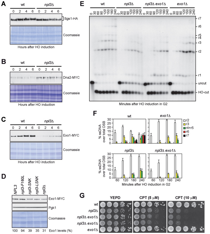
Figure 4.
NPL3 and EXO1 belong to the same resection pathway. (A–C) Exponentially growing YEPR cell cultures of strains expressing the indicated tagged proteins were transferred to YEPRG (time zero). The same amounts of protein extracts were separated on SDS-PAGE and either subjected to western blot with antibodies specific for the indicated tags or stained with Coomassie as a loading control. (D) The same amounts of protein extracts prepared from exponentially growing YEPD cultures of strains as in Figure 3, all expressing the Exo1-MYC tagged protein, were either stained with Coomassie or subjected to western blot with anti-MYC and anti-Pgk1 (loading control) antibodies. The relative intensity of the Exo1-MYC signal compared to wild type (set to 100%) was estimated after normalization to the Pgk1 band. (E and F) G2-arrested cell cultures of the indicated strains were transferred to YEPRG (time zero) in the presence of nocodazole. (E) DSB resection as described in Figure 2C. (F) Resection products in (E) were analyzed by densitometry. The mean values are represented with error bars denoting SD (n = 3). (G) Exponentially growing cell cultures of the indicated strains were spotted out onto YEPD plates with or without CPT.

