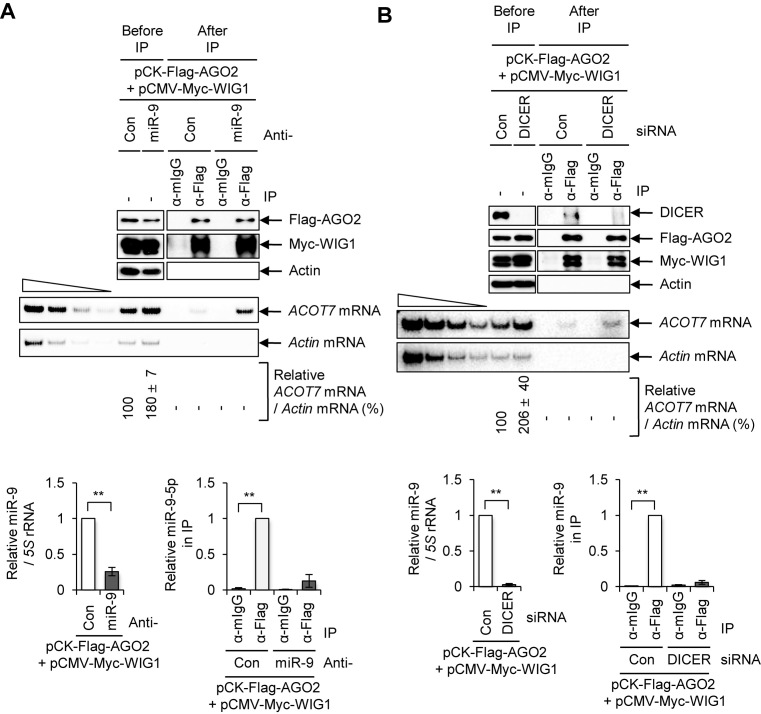
Figure 2.
Inhibition of miRNA processing and disruption of ACOT7-3΄UTR miR-9-binding site are not sufficient to block AGO2-WIG1 binding to ACOT7 mRNA. (A) Effect of anti-miR-9-5p on AGO2-WIG1 binding to ACOT7 mRNA. HEK 293T cell lysates were harvested and precipitated with anti-Flag antibody 2 days after co-transfection of pCK-Flag-AGO2 and pCMV-Myc-WIG1 plasmids in combination with anti-miR-9. From RNP-IP, precipitated proteins were subjected to Western blotting (top). RNAs in precipitates were radiolabeled with RT-PCR using ACOT7-specific primers (bottom). Actin mRNA served as a negative control. Band intensity was quantified using Quantity One software. The four leftmost lanes represent 2-fold serial dilutions of RNA, and confirm RT-PCR was semi-quantitative. Relative level of miR-9 in anti-miR-9-treated cells was normalized to that of a 5S rRNA internal control, relative to anti-miR-Con. Fold enrichment of miR-9 in Flag-AGO2 immunoprecipitates was assessed after treatment with anti-miR-9, relative to anti-miR-Con. Data are presented as means ± SD from three independent experiments. (B) Binding of AGO2-WIG1 to ACOT7 mRNA in DICER-silenced cells. Immunoprecipitation was performed with anti-Flag antibody 2 days after co-transfection of pCK-Flag-AGO2 and pCMV-Myc-WIG1 in DICER Si-transfected HEK 293T cells. Precipitated proteins were subjected to western blotting (top). RNAs in precipitates were subjected to RT-PCR using ACOT7 mRNA-specific primers (bottom). Relative level of miR-9 in DICER-treated cells was normalized to that of a 5S rRNA internal control, relative to anti-miR-Con. Fold enrichment of miR-9 in Flag-AGO2 immunoprecipitates was assessed after treatment with DICER Si, relative to anti-miR-Con. Data are presented as means ± SD from three independent experiments. *P < 0.05; **P < 0.01; #P > 0.05.

