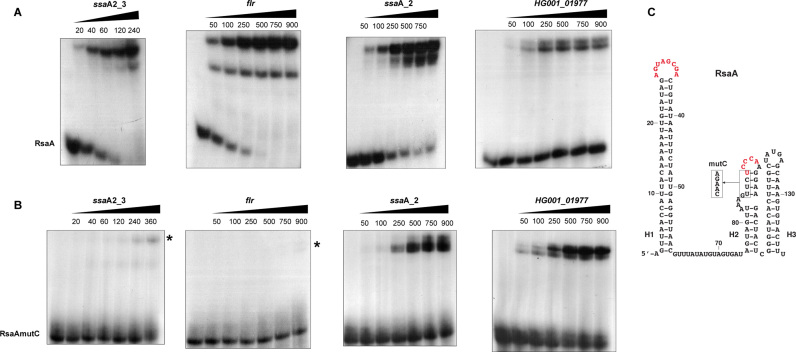
Figure 2.
Gel retardation assays to monitor RsaA binding to several mRNAs. Experiments were performed on four different mRNA targets of RsaA, which are ssaA2_3 (nucleotides −83 to +130), flr (nucleotides −47 to +120), ssaA_2 (nucleotides −128 to +125), HG001_01977 (nucleotides −390 to +122). The 5΄ end-labelled WT RsaA (A) or the mutant RsaAmutC (B) were incubated with increasing concentrations (nM) of mRNAs. In the case of flr mRNA, two complexes could be detected with WT RsaA. In the gel shift assays with RsaAmutC, residual complexes are indicated with a star. (C) Representation of RsaA secondary structure. The C-rich residues (with the mutation introduced in the RsaAmutC variant) and the second region of interaction with mgrA mRNA are shown in red.

