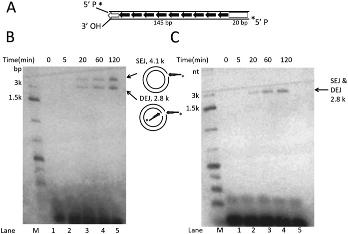
Figure 4.
In vitro analysis of Muta1 end joining with target DNA. (A) The 165-bp DNA substrate contains full length Muta1 L-TIR with 20 bp of Muta1 internal sequence. Two lines indicate the DNA double strands. On the bottom strand, the 5΄ P is radiolabeled and the 3΄ OH is exposed. (B) Reaction products on a native agarose gel reveal two bands at 4.1 and 2.8 kb, which reflect joining product SEJ (nicked circular plasmid formed by joining of one transposon end to one plasmid strand) and DEJ (linearized plasmid formed by concerted joining of two transposon ends to two plasmid strands), as indicated in the diagram on right. (C) Reaction products on a denaturing agarose gel, where only the 2.8 kb is seen, which reflects both DEJ and SEJ products.

