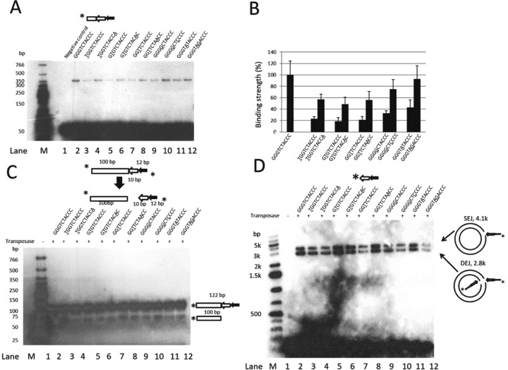
Figure 6.
In vitro analysis of the impact of the Muta1 terminal palindromic motif on DNA binding, double strand break and end joining reactions. (A) DNA binding assay. Diagram shows the structure of the DNA substrate. Underlined letters indicate mutations in the terminal motif. Reactions products are resolved on a native polyacrylamide gel. The unlabeled 165-bp DNA containing 20-bp flanking DNA and the full length Muta1 L-TIR is added in 100 (+) molar excess as competitor DNA. Fast migrating bands are the DNA substrates (∼50 bp). (B) Quantification of the DNA binding of substrates used in (A). Strength of binding was quantified with ImageJ software and repeated five times to generate the standard deviation. (C) Cleavage assay. Diagram shows the structure of DNA substrates and cleavage products. Sequences of substrates are shown; underlined letters indicate mutations in the terminal motif. Reactions products are resolved on a native polyacrylamide gel. (D) End joining assay. Diagram shows the structure of the DNA substrate. Underlined letters indicate mutations in the terminal motif. Intact pUC19 plasmid is used as target DNA and reaction products are resolved on a native agarose gel.

