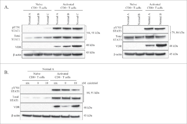
Figure 2.

Comparison of VDR and STAT levels in naïve vs. activated normal CD8+ T-cells, and the effects of calcitriol treatment on each cell type. Naïve CD8+ T-cells were isolated from 3 healthy normal donors. One half of each sample was cultured as naïve cells while the other half was activated with PHA for 24 hours. Total loaded protein per sample was 25μg, with β-actin used as a loading control. (A) Western blotting of VDR, STAT1 (pY701, total) and STAT3 (pY705, total) was performed. (B) The naïve and activated cells from Normal Donor #0 were cultured for 24 hours, with or without calcitriol treatment, lysates were probed on a protein gel blot for VDR and STAT1 (pY701, total).
