
Figure 5.
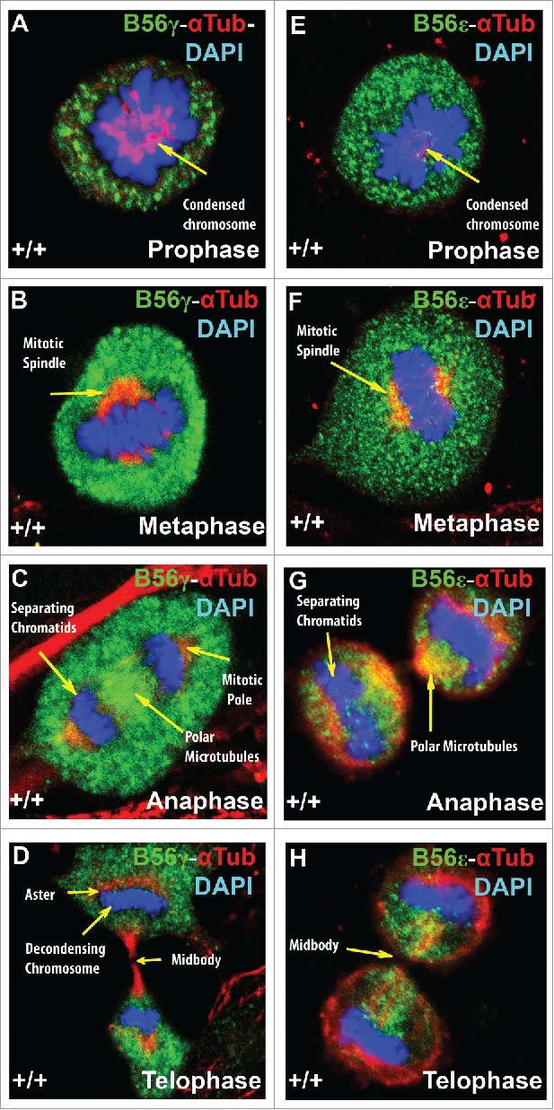
B56γ and B56ε localization during mitosis. Immunostaining of MEFs was done using antibodies against B56γ (green, A - D) or B56ε (green, E- H) along with α Tubulin (red) and DAPI (blue) in prophase, metaphase, anaphase and telophase respectively. Representative immunofluorescence images from MEFs at E14.5. Representative images from 3 independent experiments were obtained using a 60X objective.