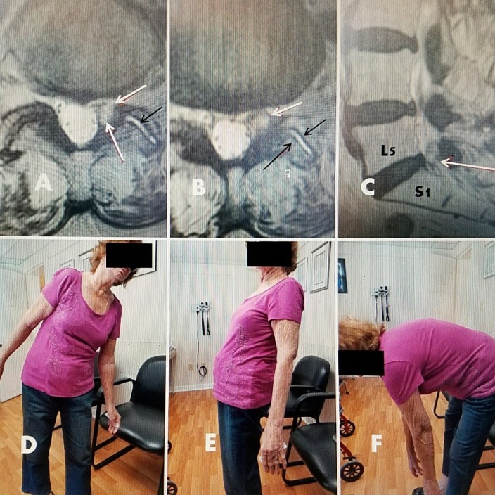
Figure 3. 79-year-old female with L5-S1 foraminal cyst with radiculopathy.
A, B: T2 axial MRI showing left foraminal facet cyst (large solid white arrow) and hyperintense fluid in left facet joint (small black arrow).
C: Sagittal T2 MRI showing posterolateral cyst at L5-S1 (white arrow).
D: Patient 18 months after drainage and ablation with lateral bending to the left, towards side of cyst.
E/F: 18 months after RF cyst ablation. Patient has recovered full movement with extension and flexion without limitation or leg pain.

