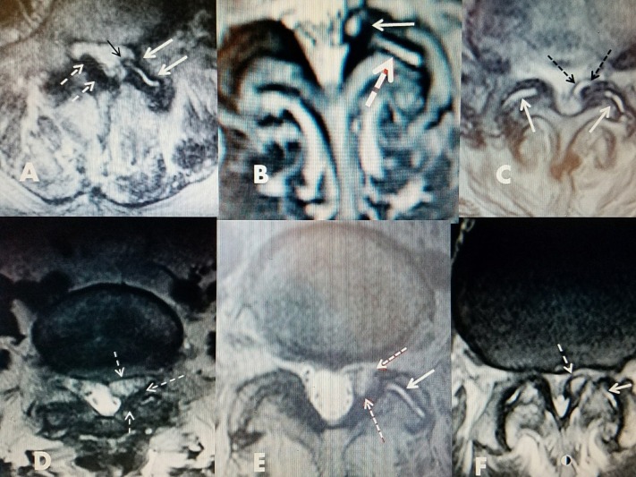
Figure 4. Axial MRI scans showing the different fluid densities within the facet cysts.
Examples of different MRI characteristics of different lumbar facet cysts.
A: Mixture of posterior ligament stenosis (dashed white arrows), development of fluid in facet joint (white arrows), and beginning of cyst on same side extending out of the ventral joint space into the lateral recess (black arrow).
B: Unilateral facet and foraminal cyst at L4-5 causing lateral recess stenosis (upper white arrow). High intensity T2 signal change indicates fluid within joint (dashed white arrow).
C: Bilateral hyperintense fluid in both facet joints (solid white arrows) shows cyst formation on one side with medial encroachment into lateral and dorsal spinal canal (dashed black arrows).
D: Foraminal cyst with more homogenous fluid on MRI that is more gelatinous (dashed white arrows).
E: Hyperintense signal of fluid in facet joint (white arrow) and more gelatinous isodense fluid in facet cyst (dashed red/white arrows).
F: Fluid in facet joint (white arrow) and bone spur from inferior facet mimicking cyst (dashed arrow).

