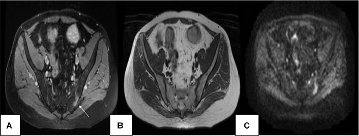
Figure 2.
MRI of SIJ: (A) axial STIR sequence (TR 2500, TE 35) shows the hyperintense signal of left SIJ (arrow); (B) axial T1-weighted sequence (TR 450, TE 21) shows the hypointense signal of left SIJ; (C) DWI (TR 2000, TE 70) shows the hyperintense signal of left SIJ with edge blurring.
Abbreviations: DWI, diffusion-weighted imaging; MRI, magnetic resonance imaging; SIJ, sacroiliac joint; STIR, short tau inversion recovery; TR, repetition time; TE, echo time.

