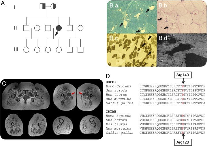
Figure. Clinical-pathologic features of patient homozygous for the HSPB1 p.Arg140Gly mutation and protein conservation between species.
(A) Family pedigree: an arrow indicates the proband; half-filled indicates distal weakness in parents who were heterozygous for p.Arg140Gly mutation. (B) Biopsy of the quadriceps muscle performed at age 27; (B.a) modified Gomori trichrome staining shows variation in fiber diameter and prominent vacuoles within many muscle fibers, arrows; (B.b) Periodic acid–Schiff preparation showed no evidence of glycogen accumulation within vacuoles (arrows); (B.c) ATPase pH 9.5 demonstrates that vacuoles are predominantly in darkly stained type 2 fibers, arrows; and (B.d) ultrastructural examination of the muscle revealed electron-dense material within vacuoles (arrows). (C) Muscle MRI demonstrating severe widespread fatty infiltration of pelvic, thigh, and calf muscles with relative sparing of the adductor longus (red arrows). (D) Conservation of HSPB1 and HSPB5 (CRYAB) amino acid sequence between species. The Arg140 residue in HSPB1 is well conserved and corresponds to the position of Arg120 in the CRYAB gene.

