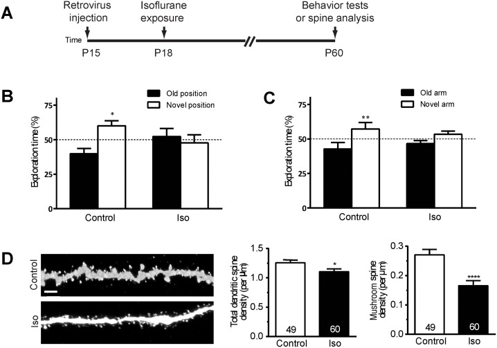
Fig 2. Isoflurane exposure impairs spatial learning and causes a loss of dendritic spines in dentate gyrus neurons.
(A) A schematic diagram of isoflurane exposure procedure for behavior tests and spine analysis. Shown in (B and C) are summaries of the object-place recognition test (B) and the Y-maze test (C) (Control n = 12, Isoflurane n = 11; **p < 0.01, Student t test). (D) Representative processed confocal images of dendritic spines of control and isoflurane-exposed green florescent protein positive (GFP) neurons at postnatal day (P) 60 (scale bar: 2 μm). Shown on right are summary plots of total and mushroom class dendritic spine density, revealing a striking loss of mature spines. Numbers associated with the bar graph indicate the number of dendritic segments examined from at least 5 mice from each group, a total of 2,586 spines in the control group and 2,818 spines in the isoflurane group were analyzed (*p < 0.05; ****p < 0.0001, Student t test). Underlying data in S1 Data under Fig 2B-D tab.

