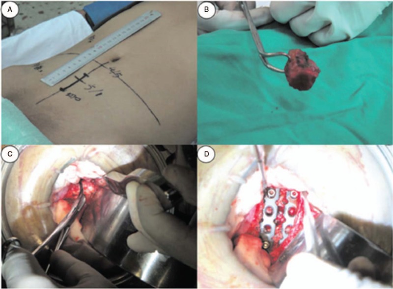
Figure 1.

Intraoperative images of a 30-year-old male. (A) A 5-cm median incision by the linea alba was made around the lesion level through the skin and along the symphysis pubis to the umbilicus. (B and C) Autologous iliac bone was packed to the prepared bone groove of the L5–S1 level. (D) Two anterior reconstructive anatomical screw-plates of suitable length were fixed anteriorly at the L5–S1 level.
