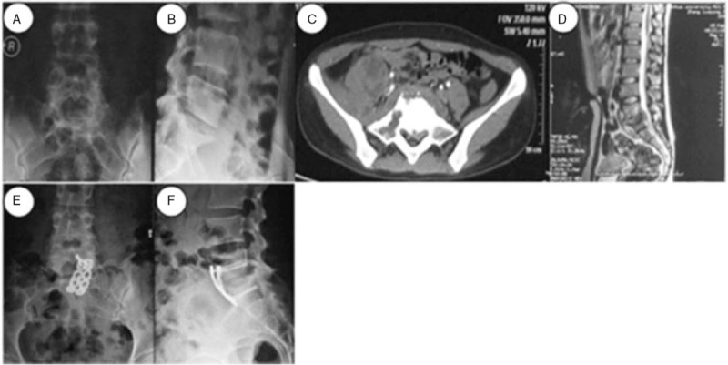
Figure 4.

A 26-year-old woman was diagnosed with L5-S1TB and underwent anterior debridement and reconstruction with anatomical screw-plate fixation. Before surgery, posteroanterior (A) and lateral (B) plain radiographs showed the L5–S1 vertebral damage with a narrowed intervertebral space and a decreased lumbosacral angle. The CT (C) and MRI (D) scans showed the destruction of L5 and S1 vertebrae, with a cold abscess compressing the neural elements. During the last follow-up visit, the x-ray images (E and F) showed a complete correction of the lumbosacral angle and a solid fusion.
