
Figure 1.
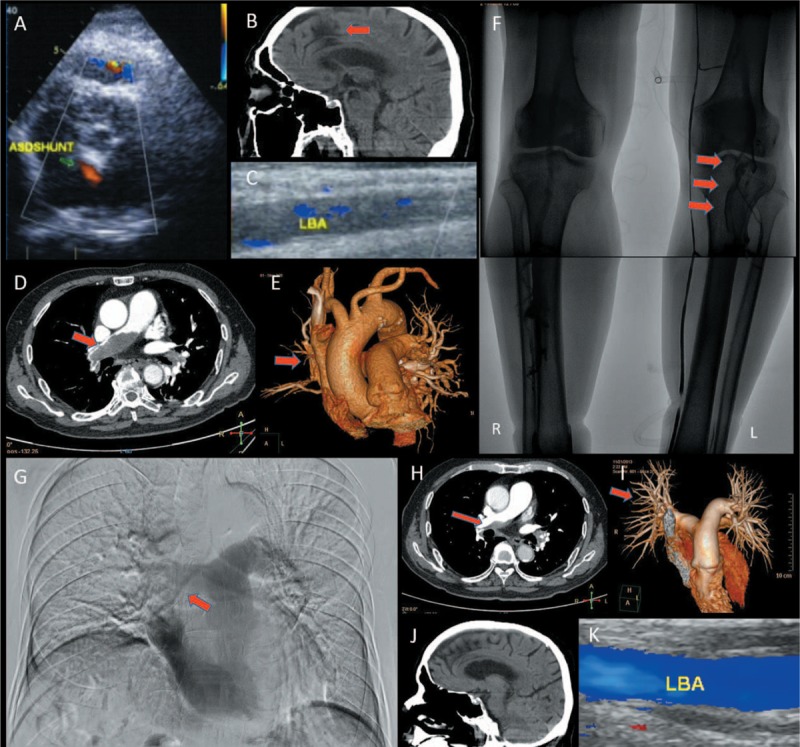
Imaging findings of case 1. (A) TTE showed an atrial septal defect complicated by atrial septal aneurysm, and a left to right shunt; (B) Head CT showed multiple lacunar infarcts, cerebral white matter demyelination, and encephalatrophy; (C) CDFI revealed total occlusion of middle-lower segment of the LBA; (D) CTPA showed extensive bilateral PE; (E) Pulmonary atelectasis was found in the middle lobe of the right lung; (F) Posterior tibial vein and anterior tibial vein were not visualized in ascending phlebography. The lower segment of peroneal vein and popliteal vein showed large filling defects (red arrow); (G) Pulmonary artery angiography showed a filling defect in the right pulmonary artery; (H, I) After 2 months of extramural hospital treatment, follow-up CTPA showed a significant improvement in the PE; (J) Head CT showing good recovery of cerebral lesions; (K) CDFI of the LBA showed good recovery.