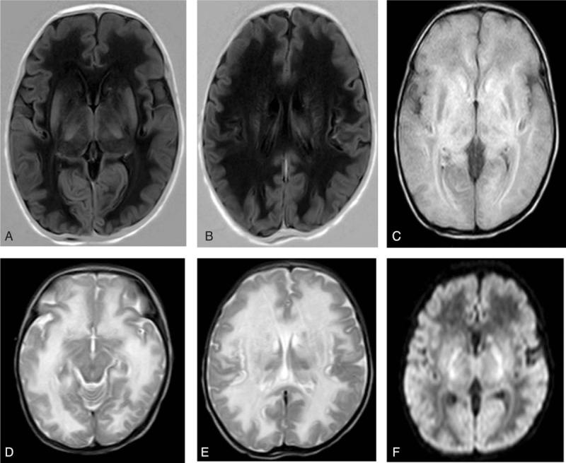
Figure 1.

Brain magnetic resonance imaging (MRI) on day 18 of life. MRI revealed extensive abnormalities in the deep white matter of the bilateral cerebral hemisphere, subcortical white matter, caudate nuclei, the dorsal thalamus, and the cerebellar hemisphere, which suggested hereditary metabolic leukoencephalopathy. (A and B) Low signal intensity on T1 weighted image (T1WI); (C) high signal intensity on fluid attenuated inversion recovery (FLAIR); (D and E) high signal intensity on T2 weighted image (T2WI); (F) high signal intensity on diffusion weighted image (DWI).
