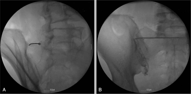
Figure 1.

Fluoroscopy-guided intra-articular pulsed radiofrequency of the left sacroiliac joint (SIJ). A, Contralateral oblique view. A 22-gauge curved-tip cannula was inserted into the wedge shape and advanced laterally and inferiorly into the SIJ. B, Antero-posterior view shows an arthrogram of the SIJ after injection of contrast material.
