Figure 1.

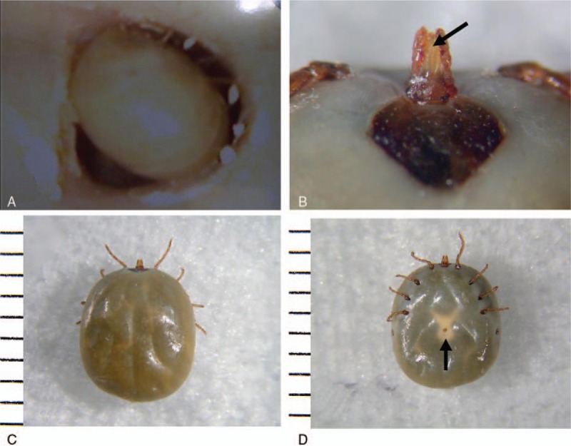
Otoscopic view of the right external auditory canal in a 12-year-old girl. (A) Although tick leg movement was confirmed, we could not see the parasite's hypostome. The extracted specimen was identified as a female A testudinarium tick (nymph stage, 6.3 × 5.3 mm); tick's (B) hypostome; (C) back; and (D) abdomen. Note the hypostome (B, arrow) and genital pore (D, arrow).
