Figure 5.
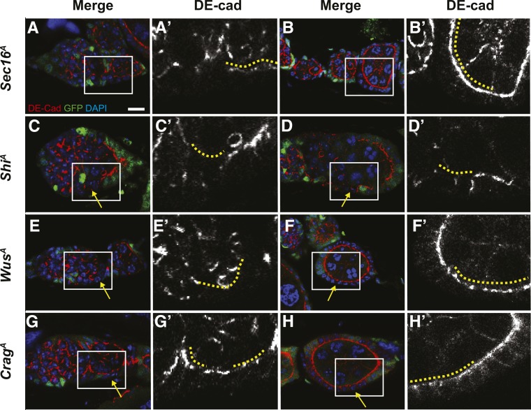
Germaria with ShiA (A-B), WusA (C-D), CragA (E-F), or Sec16A (G-H) clones stained for GFP (clonal marker, green), DE-cad (red), and DAPI (blue). DE-cad channel shown separately in A′-H′. Images of clones in the germarium shown in A, C, E, and G; images of clones in budded follicles shown in B, D, F, and H. The positions of mutant follicle cells are indicated by solid yellow lines. Boxed regions in A-H are magnified in A′-H′. Yellow arrows indicate GFP− homozygous mutant follicle cells. DE-cad is present on the apical membranes of heterozygous, GFP+ follicle cells in all cases but absent from the apical domains of ShiA follicle cells in the germarium and in budded follicles; absent from the apical domains of WusA follicle cells in the germarium and disrupted in WusA follicle cells of budded follicles; present but mildly disrupted in CragA and Sec16A follicle cells in the germarium; and normally localized in CragA and Sec16A follicle cells in budded follicles.

