Figure 7.
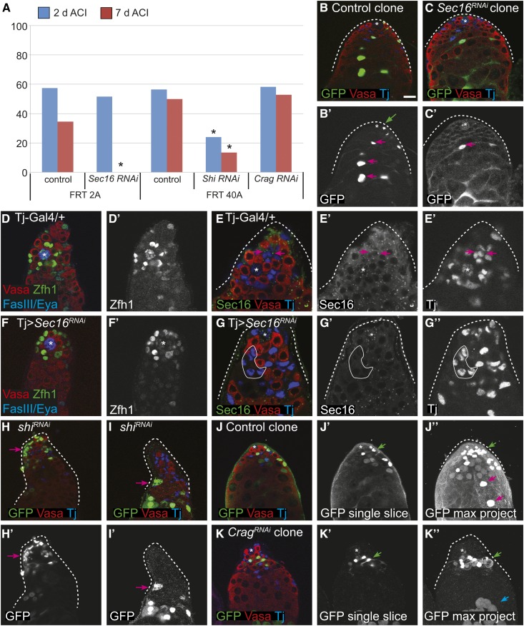
(A) CySC niche competition assays for control and RNAi knockdown of the indicated genes and matched FRT control clones at 2 (blue bars) and 7 (red bars) days ACI. * P < 0.05 compared to the control in the same set. (B and C) Control clones with CySCs [(B′), green ←] and differentiating daughter cells [(B′), pink ←] were recovered at 7 days ACI; whereas Sec16 RNAi CySCs were not recovered at 7 days ACI, but their differentiating daughter cells were observed [(C′), pink ←]. (D–G) Depletion of Sec16 from the entire somatic lineage does not perturb the stem cell niche as a Tj > Sec16 RNAi testis (F and G) looks very similar to a control testis (D and E) in terms of the number of CySCs (Zfh1-positive cells). (D′ and F′) Zfh1 channel only. (G′-G′′) Sec16 and Tj channels only. Sec16 localizes to discrete cytoplasmic puncta in cyst cells of wild-type testes (E–E′), and this signal is eliminated in a Tj > Sec16 RNAi testis [(G), representative cyst cells outlined with a white line]. (H and I) shi RNAi CySC clones have abnormal phenotypes, including pronounced fragmentation that suggests the cells are dying [(H′), ←] and aberrant membrane formations that suggest incomplete germ cell ensheathment [(I′), ←]. (J and K) In a testis with control clones at 7 days ACI, a labeled CySC [(J′′), green ←] and labeled differentiating cyst cells [(J), pink ←] were readily observed. In contrast, in Crag RNAi clones, labeled CySCs were readily observed [(K′′), green ←] but differentiating Crag-depleted cyst cells were not seen. Note that the GFP cells at the bottom are labeled germ cells [(K′′), blue ←]. (J, J′, K, K′) are single confocal sections whereas (K′′) and (J′′) are maximum projections.

