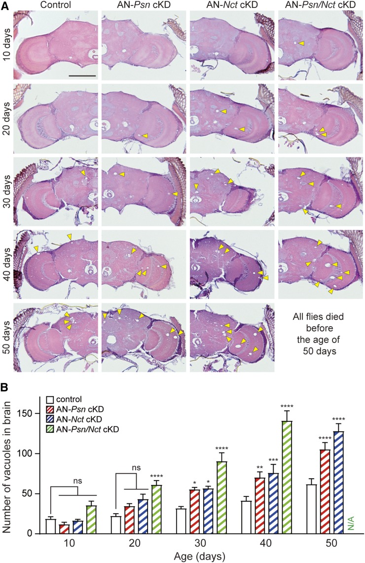
Figure 8.
Age-dependent neurodegeneration in adult neuron-specific Psn and Nct cKD flies. (A) H&E staining in frontal brain sections revealed age-dependent neurodegeneration. Increased number of vacuoles (marked by arrowheads) indicates neurodegeneration in the brain. Bar, 100 μm. (B) Quantification of vacuoles in the brain of control (elav-Gal4/y;; tub-Gal80ts/+, black), AN-Psn cKD (elav-Gal4/y;; tub-Gal80ts/UAS-shPsn2, red), AN-Nct cKD (elav-Gal4/y;; tub-Gal80ts/UAS-shNct2, blue), and AN-Psn/Nct cKD flies (elav-Gal4/y;; tub-Gal80ts, UAS-shPsn2/UAS-shNct2, green). Neurodegeneration is minimal in the brain of control flies. The brains of AN-Psn cKD and -Nct cKD flies show significant age-dependent neurodegeneration compared to brains of control flies at the age of 30, 40, and 50 days. The brains of AN-Psn/Nct cKD flies show significant age-dependent neurodegeneration compared to the brains of control flies at the age of 20, 30, and 40 days. All AN-Psn/Nct cKD flies died before the age of 50 days. Total number of vacuoles >5 μm in diameter was counted throughout all sections of entire brains. n = 10–17 brains per genotype. All data are expressed as mean ± SEM. Statistical analysis was performed using two-way ANOVA with Dunnett’s post hoc comparisons. Two-way ANOVA showed main effects of genotype [F(3, 197) = 22.77; P < 0.0001] and age [F(4, 197) = 57.92; P < 0.0001], with an interaction between these factors [F(12, 197) = 19.94; P < 0.0001]; ns, nonsignificant; *P < 0.05, **P < 0.01, ****P < 0.001, ****P < 0.0001.

