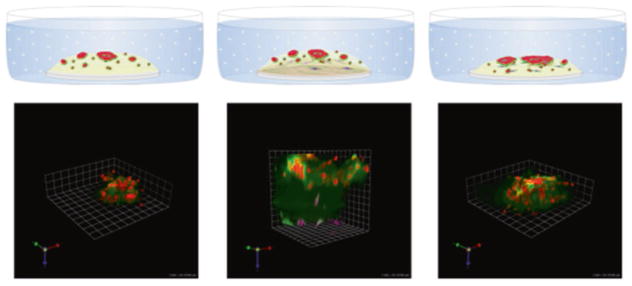
Fig. 1.
Pathomimetic avatars: schematic diagrams and representative images of 8-day cultures. Left: MDA-MB-231 human breast carcinoma cells in reconstituted basement membrane (rBM) + 2% rBM overlay. Middle: 231 cells in a top layer of reconstituted basement membrane (rBM) overlaid with 2% rBM and WS12Ti human breast carcinoma-associated fibroblasts embedded in a lower layer of collagen I. Right: tumor cells and fibroblasts mixed and plated together in rBM and overlaid with 2% rBM. Quenched fluorescent protein substrates (DQ-collagens IV and I) are mixed with rBM and collagen I, respectively. Note the more extensive degradation and increased size of 231 structures in the presence of fibroblasts. Red, magenta, and green represent 231 tumor cells, fibroblasts, and fluorescent cleavage products of the substrates, respectively

