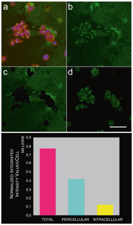
Fig. 4.
Quantification of degradation of DQ-collagen IV by pathomimetic avatars of HCT116 human colon carcinoma cells. (a) Single optical section of 3D pathomimetic avatar at equatorial plane showing fluorescence of cells (magenta) used for cytoplasmic binarized mask, nuclei (blue) used for counting cells, and degraded DQ-collagen IV (green). (b) Total degraded DQ-collagen IV in this optical section. (c) Pericellular degraded DQ-collagen IV in this section. (d) Intracellular degraded DQ-collagen IV in this section. Quantification of degraded DQ-collagen IV is done in each optical section of 3D volume, totaled and normalized to the number of cells. Image arithmetic is used to separate total proteolysis (magenta bar) into intracellular (yellow bar) and pericellular (cyan bar) components

