Fig. 8.
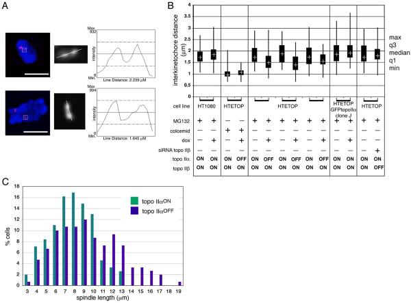
The effect of topo IIα depletion on metaphase inter-kinetochore distance. (A) Distances between sister centromeres of human chromosome 11 detected by FISH using D11Z1 DNA as a probe were assessed using Leica Deblur software. Scale bar, 10 μm. (B) Measurements made of treated cells in parallel with untreated controls (treated/ untreated pairs indicated by brackets) are presented as a boxplot showing the third and first quartiles, with the median indicated by a cross in each box. The maximum and minimum values are indicated by the ends of the vertical lines. Each plot is based on ≥40 measurements. In HTETOP topo IIα was depleted by 72 hours doxycycline exposure. Cells were arrested using MG132 (10 μM for 2 hours 30 minutes) allowing distances under tension to be measured. For the doxycycline-treated/ topo IIα-depleted HTETOP cells data from 3 independent experiments is presented. In each case the decrease in the distance across the centromere domain under tension following topo IIα-depletion is significant (p = 0.001, 0.0001, 0.01 respectively). To deplete topo IIβ HTETOP cells were transiently transfected with siRNA (72 hours). Colcemid-treated HTETOP cells served as a control where no spindle or tension existed. The lack of any effect from doxycycline-treatment itself was confirmed by analysis of MG132-arrested parental HT1080 cells. Clone J is a derivative of HTETOP that constitutively expresses topo IIα as a fusion with the C terminus of eGFP (Carpenter and Porter, 2004). (C) Quantification of spindle lengths in HTETOP cells arrested using MG132 (10 μM for 2 hours 30 minutes) following 0 (topo IIαON) or 72 hours (topo IIαOFF) doxycycline exposure. Measurements of pole-to-pole distance, based in γ-tubulin and DAPI staining, were collected from 3 independent experiments (topo IIαONn154, topo IIαOFF n151).

