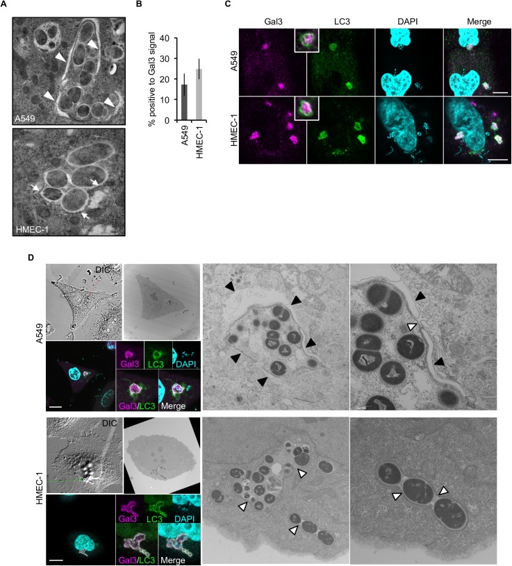
Fig 2. Endothelial cells are intrinsically defective in formation of double-membrane structures surrounding GAS.
(A) Representative EM images show double-membrane structure (arrowheads) in A549 cells, but not in HMEC-1 cells. HMEC-1 cells were only positive for single membranes (arrow) at 1 h post-infection. (B) GAS with ruptured endosomal membranes in A549 and HMEC-1 cells were stained by anti-Gal3 antibody. Error bars indicate SD of three independent experiments. (C) Immunocytostaining images show LC3 signals on GAS with ruptured endosomal membranes (Gal3) at 1 h post-infection. Scale bar, 10 μm. (D) Representative CLEM images show LC3 (GFP) and Gal3 (Strawberry) signal on membrane structures surrounding GAS. A total of 16 (epithelial cells) and 36 (endothelial cells) GAS-containing vacuoles doubly positive for Gal3 and LC3 were observed. Black arrowheads double-membrane structure; white arrowheads, single-membrane structure. See also S2 Fig. Scale bar, 10 μm.

