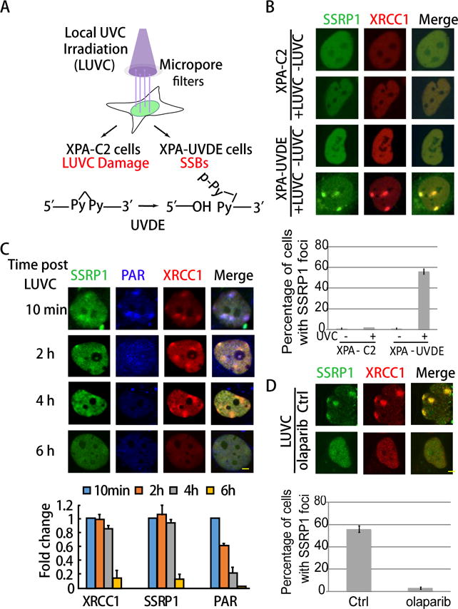
Figure 2. FACT is enriched at SSBs together with XRCC1 at damage sites after PAR degradation.

A. Scheme of the assay used to induce specific SSBs in XPA-UVDE cells after local UVC irradiation. XPA-UVDE/C2 cells were exposed to 254 nm UVC light through a polycarbonate isopore member filter. After local UVC (LUVC) irradiation, UV damage was produced and unrepaired in both XPA-C2 and XPA-UVDE cells. In XPA-UVDE cells, SSBs were introduced by UV damage endonuclease (UVDE) at the unrepaired UV damage sites. B. Recruitment of GFP-SSRP1 at SSBs produced in XPA-UVDE cells after 100 J/m2 local UVC irradiation. No enrichment of SSRP1 was observed in XPA-C2 cells. Quantification of percentage of foci positive cells of SSRP1 is shown. Cells showing > 3 foci were defined as positive cells. The error bar represents three independent experiments with 50 cells in each. C. Staining of pAR in GFP-tagged FACT and RFP-XRCC1 expressing XPA-UVDE cells at 10 min, 2 h, 4 h, and 6 h after 100 J/m2 LUVC irradiation. Percentage of foci positive cells of PAR, FACT and XRCC1 in XPA-UVDE cells at 10 min is normalized as 100%, fold decrease of foci positive cells at 2 h, 4 h, and 6 h after 100 J/m2 LUVC irradiation relative to 10 min foci formation is shown. The PAR signal disappeared while FACT and XRCC1 were retained at SSBs 2 h after damage production. D. Co-localization of GFP-tagged SSRP1 and RFP-tagged XRCC1 w/o PARPi. Quantification of percentage of foci positive cells of SSRP1 is shown. Cells showing > 3 foci were defined as positive cells. The error bar represents three independent experiments with 50 cells in each.
